 搜索
搜索

热门供应商
该产品暂无相关的供应商
PK的排行更多
- 台湾路昌 振动...
- 日本理音 测振仪...
- 日本理音 激...
- 美国康塔仪器 ...
- 北京拓必拓 微...
- 上海舜宇恒平 在...
- 荷兰Skalar...
- 德国德尔格 ...
- 北京拓必拓 超...
- 日本Kanoma...
最新更新的同类仪器更多
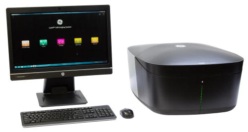
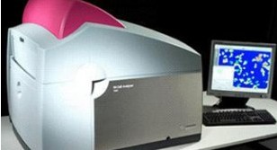


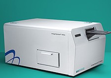
最新产品 > 生物分析仪器 > 其他生物分析仪器 > 细胞成像系统>Molecular Devices 高内涵细...
更新时间:2026-06-14 09:26:10 点击:531次
| 产品型号 | ImageXpress Velos |
|---|---|
| 参考报价 | 返回顶部 面议 |
| 基本规格 | |
| 产品类型 | 返回顶部 细胞成像系统 |
| 品牌 | 返回顶部 Molecular Devices |
| 产地 | 返回顶部 美国 |
| 返回顶部 我来纠错(有奖) | |
| 技术参数 | |
| 仪器重量 | 返回顶部 140(lbs)59(kg) |
| 仪器尺寸 | 返回顶部 W(in):18.5 D(in):29 H(in):15W(cm):47 D(cm):73.7 H(cm):38.1 |
| 其他信息 | |
| 产品简介 |
Experience the benefit of multiplexed, cell- and bead-based assays performed during high content screening in a high throughput screening environment, ensuring heterogeneous responses can be captured earlier in the research and discovery of new therapeutics. Now multiplexed, homogeneous, bead and cell-based assays become easy to develop and run as robust primary screens. The cytometer can scan a plate in as little as 3 minutes, regardless of cell- or well-density while resolving objects at micron resolution in four colors simultaneously. These fast scan times and whole well analysis can gather data from >150,000 wells per 8-hour day, achieving the acquisition rates expected of high-throughput screening. The cytometer‘s speed comes from unique design elements including a large depth of focus and large field of view which eliminates the requirements to periodically focus or acquire multiple exposures to cover an entire field of view. A light scatter (label free) mode allows generation of data from non-fluorescent objects (e.g., stem cell colonies where addition of dyes is unacceptable). To help simplify the workflow of moving suspension cytometry assays into the screening environment, the ImageXpress® Velos Cytometer leverages a microplate-based environment to streamline evaluation of both adherent and suspension cell types, eliminating separation steps for adherent cells or colonies while eradicating daily routine procedures to prime and clean fluid lines. This well-based environment utilizes small aliquots (384- or 1536-well plates), preserving valuable stem or primary cells. See the poster "High-Throughput Multiplexed Assay for Analysis of Hematopoietic Stem Cells Differentiation and Hematopoietic Toxicity". Additionally, a microplate based assay allows samples to be rescanned to confirm results or gather additional content at a higher magnification on other systems. The ImageXpress Velos Cytometer is ideally suited for capturing rare events from a variety of heterogeneous samples including large volumes of beads or cells, cell colonies, tissue samples, or even micro-array slides. A wide range of fluorophores can be accommodated on the cytometer giving the user flexibility in optimizing the multiplexing of their assays. This is complemented with the ability to detect low levels of fluorophore in homogeneous no wash assays. Users are not limited to making fluorescent intensity measurements, but can also make solution or object-based anisotropy (fluorescence polarization) experiments. "On-the-fly" image analysis provides immediate cellular results to detect signals above a user-specified background. Size, shape, and brightness are among the parameters used to define objects of interest (cells, colonies, or beads) while debris or other inappropriate objects are ignored. This is complimented by De Novo Software‘s FCS Express 4 Image Cytometry Software to analyze trends within heterogeneous or large cell populations so you can bring additional resolution to your image-based cytometry assays. Specifications The ImageXpress Velos SL Cytometer is a single-laser system configured from the selection of laser lines offered. The ImageXpress Velos DL Cytometer is a dual-line laser system consisting of a 488 nm laser system plus one additional laser line from the selection of laser lines offered. Note the ImageXpress Velos Cytometer is a Class I Laser Product. Laser lines offered and selectable through software 405 nm 440 nm 488 nm 532 nm 640 nm User selectable detection modes via software Four independently adjustable photomultiplier tubes Fluorescence 4 color simultaneous Anisotropy / Fluorescence Polarization 2 color simultaneous Laser scatter Space and weight Specifications: W(in):18.5 D(in):29 H(in):15 Weight(lbs):140 W(cm):47 D(cm):73.7 H(cm):38.1 Weight(kg):59 in = inches, lbs = pounds, cm = centimeters, Kg = Kilograms The ImageXpress Velos Cytometer is covered under multiple United States and international pending patents. 返回顶部 |
| 应用文章 | 返回顶部 此仪器尚未有相关文章 |
| 产品样本 | 此仪器尚未有相关样本 返回顶部 |
| 应用范围 | 返回顶部Common assays include: • Apoptosis • Cell cycle • Cell proliferation • Cell Migration or Invasion • Cytotoxicity • Cell- or bead-based homogeneous assays • Hybridoma screening • Adherent cell cytometry • Single cell phospho-protein detection • Protein-protein interactions in living cells • Spot- and bead-based arrays in plates • Stem cell or primary cell surface marker expression • Toxicity in model organisms |
| 用户数 | 返回顶部 此仪器尚未有用户数 |
| 技术创新 | The ImageXpress Velos is extremelyfast and thus moves HCS into the high throughput screening arena. It can scan and simultaneously analyze objects plated on any clear bottom microplate in <3 minutes. With automation it can be configured to acquire in excess of 150,000 wells in an 8 hour day, faster than any other HCS or cytometer now on the market. Whole well scanning gathers data from large fields-of-view and sufficient objects for statistical validity. Reduces the number of cells used as compared to standard microplate readers. Large depth of field (+/- 200 microns) eliminates focus steps during acquisition of any clear bottom plates (including tissue culture plates) as well as easy imaging of large objects such as 3D cell cultures, mammospheres or zebrafish embryos. Patented confined detection region reduces background from out of focus fluorescence and enables homogeneous / no-wash fluorescence assays like cell surface marker detection (e.g., E-cadherin), fluorescent ligand-receptor interactions, or antibody discovery. Anisotropy detection mode enables solution and bead fluorescent polarization assays as well as simplified live cell FRET assays. To learn more about Anisotropy and live cell FRET by Anisotropy click here. Choose from 5 laser lines (405, 440, 488, 532, and 640 nm) to optimally excite fluorophores required for your application. Photo-multiplier tubes provide sensitive and simultaneous detection of 4 emission channels in the intensity domain and two in the anisotropy domain enabling multiplexed assays, for example running mitotic index with apoptosis. Learn More about 3 color mitosis and apoptosis assays. Laser scatter mode for label-free detection of cells when intervening with dyes is not acceptable, for example in quality control of sterile cell cultures. Microplate-based environment streamlines evaluation of both adherent and suspension cells and eliminates separation steps for adherent cells or colonies often required for flow cytometry or other non-plate based systems. Acquire from any clear bottom plate, including tissue culture plates, for easy quality control of cells that need to be returned to incubator and kept sterile. Small footprint for easy benchtop installation when space is at a premium. Adjustable sampling frequency allows data to be acquired at 10, 5, and 2.5 microns. High sampling frequency helps to indentify small nuclei, neurons, or bacteria, or acquire and analyze a plate in as fast at 30 seconds (10 x 40 microns) when time is a premium. Learn more about extremely fast solution based fluorescent polarization assays. On-the-fly analysis enables users to simultaneously acquire, count, and measure objects so that results are available at the end of each scan. Robust, simple, and fast object detection means no microscopic imaging experience is required for setup making it easy for anyone in the lab to establish and run assays. Cell-by-cell and well-by-well results of object intensity, area, and shape are generated during each scan, allowing easy classification and gating of objects to streamline your work flow. Output data in a variety of formats (.csv, .txt, .fcs) for easy downstream analysis and manipulation in a variety of software packages including FACS software. MDCStore™ Datamanager compatible files facilitate analysis with MetaXpress and AcuityXpress Software for quantifying objects like zebrafish embryos or enterprise level data visualization and trend analysis. Dedicated support team of service engineers, field applications specialists, and in-house Ph.D. level technical scientists available to install your cytometer, train, and answer your questions so that you can get to your experiments quickly. 返回顶部 |
| 最新动态 | 返回顶部 此仪器尚未有最新动态 |
| 相关视频 | 返回顶部 此仪器尚未有相关视频 |
| 相关耗材和配件 | 此仪器尚未有相关耗材和配件 返回顶部 |
| 相关仪器 | 返回顶部 此仪器尚未有相关仪器 |
| 网友评分 |
评分:
4星:0人 3星:0人 2星:0人 1星:0人 返回顶部 |
|
该产品暂无相关的供应商 |


暂无评论
- 网友评论:
- 已有0条评论